
Main objective of the project is to develop a multi-modal medical imaging technology that reduces the use of ionizing radiation to the minim possible level. This technology should operate in real time for acquisition as well as for tomographic reconstruction in realistic diagnosis and intervention scenarios, like an operating theater.
Specific objectives are:
- Development of a high-resolution ultrasound imaging technique combining reflectivity and tomography methods, adapted for real-time operation.
- Design of radiation detectors compatible with positioning systems for its application in surgical environments (surgeries or biopsies). Use of these techniques in surgical X-Ray arcs.
- Development of a real-time technology for data fusion between multi-modal images with spatial localization into the intervention scenario.
- Design and implementation of ultra-fast algorithms for tomographic reconstruction.
GSTU group from CSIC worked with UPM and UAH groups in the fist objective: To develop a high-resolution ultrasound imaging and tomography system, specifically adapted for early detection of breast cancer.
Multi-modal capacity of ultrasound imaging was investigated, and three complementary techniques were implemented:
- Circular compounded ultrasound image with higher resolution and contrast than conventional echography (Fig. 1a).
- Speed of sound ultrasound tomography, which gives greater specificity for malignant cysts (Fig.1b).
- Phase coherence imaging, which eliminates artifacts and improves detection of micro-calcifications.
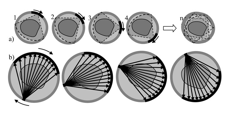
Fig. 1- Multi-modal ultrasound imaging concepts: a) Circular compounding of reflectivity images, one array in pulse-echo, 360º; b) Speed of sound tomography, with two opposite arrays.
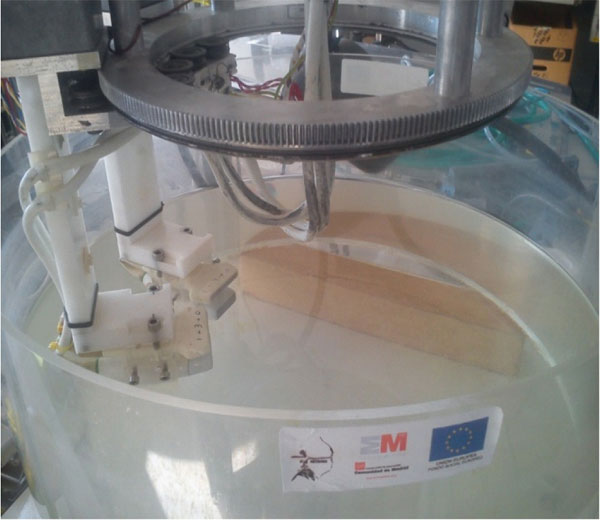
Experimental work has been carried out in a custom developed prototype, with which tissue mimicking phantoms were imaged with the abovementioned methods. Two arrays with 128 elements and a central frequency of 3.5 MHz can be precisely positioned around 360º with software controlled stepper motors (Fig. 2). The tissue mimicking phantoms (a commercial breast phantom and custom designed agar-gel phantoms) were placed in the center of the inspection tank.
An important part of the project was to numerically characterize the circular compounded image with regard to resolution, sensitivity and homogeneity, because no closed formulation has been previously described in literature (Fig. 3). An interesting result was that resolution, both in the radial and in the tangential directions, is practically constant and similar to the axial resolution of each individual image. Furthermore, it was found that 16 to 20 individual images are enough to obtain a high-quality compounded image, and increasing that figure gives no appreciable improvement in image quality.

Fig. 3- Left: Radial resolution with variations between 0.19 y 0.23 mm; Right: Tangential resolution, with variations between 0.24 y 0.3 mm. Total Diameter = 170 m. Color scale in mm
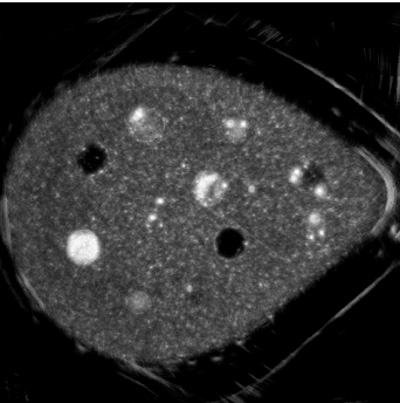
Figure 4.left shows a compounded image of a breast phantom obtained with the ARTEMIS project methodology. Solid masses and anechoic cysts are clearly seen, with good resolution and contrast. On Fig. 4.Right, a Phase Coherence Imaging of the central region is shown, in which micro-calcifications can be better evaluated.
 |
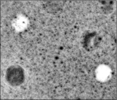 |
| Fig. 4. (Left) Circular Compounded image of a breast phantom (Right) Detail of the central region after applying the Phase Coherence Technique. | |
Speed of sound maps have been obtained by signal acquisition in through-transmission mode with opposite arrays. Based on the time-of-flight information registered from different angular positions, tomography reconstruction algorithms were implemented to obtain the velocity map. Research work has been carried out in the optimization of the reconstruction algorithm as well as in the implementation of a robust sound propagation simulator. Several algorithms were tested, from simpler ones based on ray-tracing methods (Fig. 5) to more complex ones based on full-wave iterative solutions. (Fig. 6).
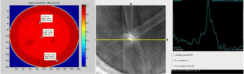
Fig.5- Image reconstructed with 2D-OSEM algorithm in an agar gel phantom.
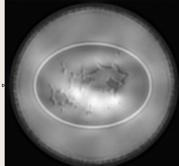
Fig.6- Full-wave reconstruction with a numerical phantom.
In view of the promising results, the collaboration between the consortium members during this project will continue after its completion. It was concluded that these imaging modalities could be very useful for the early detection of breast cancer noninvasively. Following this line, a new project proposal was presented in which ultrasound image is combined with functional PET image to improve cancer detection.
For clinical application of the proposed technique, it is necessary to design and manufacture a full ring array and a high channel count electronic to reduce scanning time. Within this project, modular system architecture with up to 2048 channels was defined, and first prototypes of the main system blocks were manufactured and tested. The final system will allow acquiring a 3D image of the whole breast volume in less than 1 minute.
 |
 |
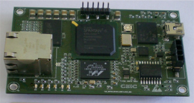
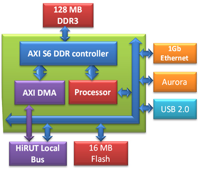
| Fig. 7-Interface and control unit based on Spartan-6 FPGA family: 1GB DDR-3, USB-2.0 and GigaEthernet |
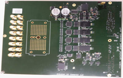
Fig. 8- First prototype of a 32 channel ultrasound module. |
CSIC and UAH groups have developed the main elements of the new architecture: the Interface and control unit, UCI (Fig. 7), and a first prototype of a 32 parallel channel pulser-receiver board (Fig.8), using state of the art electronics and most advanced PCB design techniques.
The UCI was designed as a System-On-Chip based on an embedded RISC processor, and it is responsible of sequencing the system operations and to transfer the results to a PC application (Fig. 9). For Ethernet communications, a mixed solution based on TCP and UDP was adopted. For the USB connection, a custom developed DMA block sends and receives data to the external DDR3 chips without intervention of the embedded processor.
For high-speed data transfer between the ultrasound system and the PC, a solution based on the AURORA protocol over 2.4 Gbps MGTs lines was developed. A fiber optic physical layer was used, and sustained rates of 210 MBytes/s were achieved, one order of magnitude faster than with USB 2.0 protocol. PCIe protocol was used in the PC side.
 |
 |
| Fig. 9- Internal architecture of the SoC for the UCI module. | Fig. 10- Comunicaciones multigigabit basadas en MGTs |
A vhdl IP core was developed to control the Analog Front-End -AFE- of the system, including precision delayed excitation (resolution of +2.5 ns) and signal acquisition at 64 MSPS and 14 bits. Automatic synchronization and bit-alignment algorithms were developed to handle the 0.5 Gbps links between the AFE and the FPGA.
A custom solution was developed for accessing the external DDR3 memory bank from different agents inside de SoC -processor, HW accelerator, USB, etc-. FPGA resource efficient and high-performance DDR memory controllers were also designed. During the project, High Level Synthesis -HLS- methodologies were used for the design of the FPGA cores. In particular, a distributed architecture for a digital scan converter was proposed and tested.
J. Camacho, L. Medina, J.F. Cruza, J.M. Moreno, C. Fritsch, “Multimodal ultrasonic imaging for breast cancer detection”, Archives of Acoustics, Volumen 37-3, Páginas 253-260, Setiembre de 2012, DOI: 10.2478/v10168-012-0033-4.
J.M. Moreno, M. Sánchez, J.F. Cruza, L. Medina, “Mini-Módulo HW de altas prestaciones, basado en Saprtan6 con análisis de integridad de señal”, Actas de las XII Jornadas de Computación Reconfigurable y Aplicaciones (JCRA), pp. 4-9, ISBN:978-84-695-4470-9, 2012.
J. Camacho, C. Fritsch, “Phase coherence imaging of grained materials”, IEEE Trans. on Ultrasonics, Ferroelectrics and Frequency Control, Volumen 58-5 , Páginas 1006-1015, Mayo de 2011, DOI: 10.1109/TUFFC.2011.1901
Camacho, Jorge; Brizuela, Jose; Moreno, Jose Miguel; F. Cruza, Jorge; Fritsch, Carlos “Instrumentación para tomografía ultrasónica médica y primeras imágenes”, Conferencias y Comunicaciones TECNIACUSTICA'11, ISBN: 978-84-87985-20-1, 2011.
Foundation for Biomedical Research of the Gergorio Marañón Hospital, Universidad Politécnica de Madrid, Universidad Complutense de Madrid, Spanish National Research Council, Universidad de Alcalá