
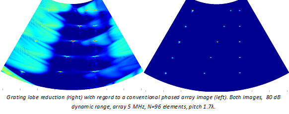
The project aims to the development of theoretical and practical support for a new technique devised by our group, called Phase Coherence Processing (PCP). It addresses the improvement of the quality of ultrasound images obtained with phased array technology. The PCP technique increases the lateral resolution and, simultaneously, suppresses grating and sidelobe indications.
These joint characteristics have been obtained for the first time. The performance has been preliminary demonstrated by simulation and real experiments. Dramatic improvements in the image dynamic range (40 dB) and in the lateral resolution (x2) have been obtained. These results may have an important impact in the medical (echography) and industrial (NDT) fields.
Also, they open a new research line with many possible applications, whose exploration is the main objective of this project. Three aspects will be addressed: a) Theoretical assessment of the PCP technique, b) Real-time implementation and c) An advanced application.
The first part will focus on different topics to assess the limits of the new technique and to evaluate its performance for new applications. The hardware implementation of the PCP technique and its integration with a phased array architecture will allow the operation in real-time. Finally, the application of the technique to breast cancer detection is proposed. This choice is based on the improvements in contrast and resolution provided by PCP, which might lead to a substitution of current practices (mammography, based on X-rays) by ultrasonic exploration.
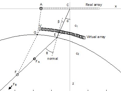
A new method to perform dynamic depth focusing through interfaces by means of an intermediate virtual array

New real-time dynamic depth focusing architecture
When a coupling medium is inserted between the array probe and the inspected body, focal law computing becomes complicated by the refraction effects at the interface. Furthermore, there were not previously known techniques to perform dynamic focusing by hardware in these conditions.
This project addressed the problem following a two step procedure: First, a virtual array that operates in a single medium with nearly equivalent time-of-flight to the foci is obtained. Then, a simple hardware was devised to perform dynamic focusing in real-time. The circuit operates with arrays of any geometry as required by the virtual array in presence of arbitrarily shaped interfaces.
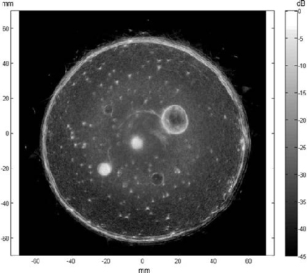
A breast phantom ultrasound compounded image showing masses, cysts and micro-calcifications using the new dynamic depth focusing procedures.
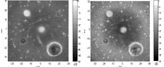
Detail of the central part of the breast phantom image: left: compounded image; right: phase coherence processed with removed artifacts and improved micro-calcifications resolution